Dental Archives Based on Images
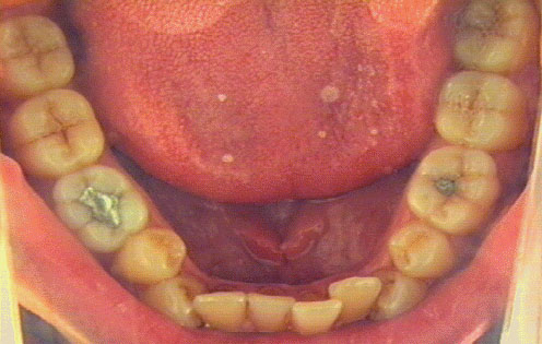
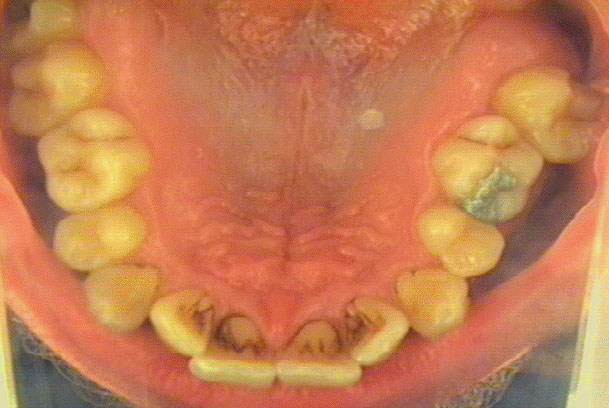
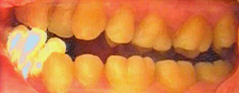
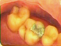
The Orthoscope is an equipment for
acquisition, processing, and archiving of images of patients
mouth or skin. The Orthoscope can capture and process images of a
single tooth, a group of teeth, or the whole dental arc. A
dentist can easily (1) observe the situation in the mouth, (2)
demonstrate the intended plan of a treatment to the patient, and
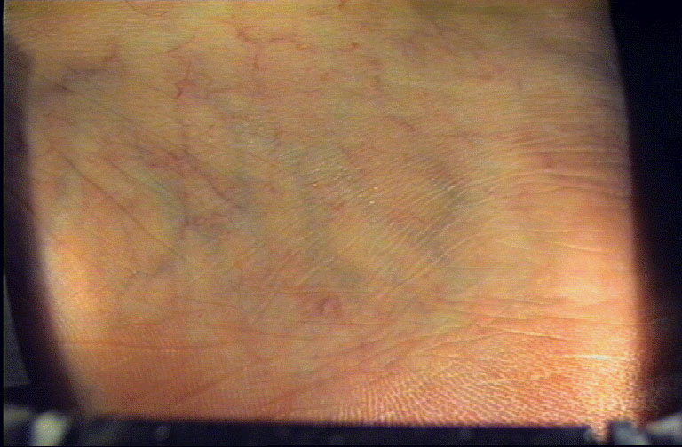
(3) document the treatment results. A dermatologist can evaluate
treatment progress. Unlike other methods, our device shows
geometrically undistorted calibrated image, and capture the whole
teeth arc in one image.
New images.
The Orthoscope should be used in daily practice. The image
processing module is connected to an insurance office system and
medical archives. This eliminates time consuming literal
description of the patient dental/dermatological status. The
images can be used later for checking of the diagnosis and the
treatment.
Features:
- Device for capturing of color images in dentistry and
dermatology.
- Geometrically undistorted calibrated images.
- Measurement of distances and areas.
- Image analysis functions.
- Archiving of captured images.
- Comparison of archived and new images supported by
overlapping function.
- Client-server architecture with interface to
administrative database.
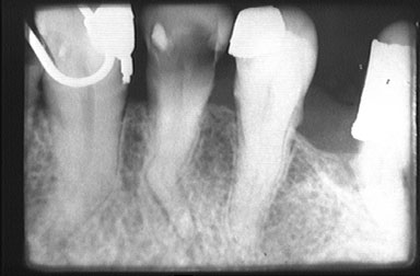
- Accessory for capturing X-ray images from films.
- Import/export of images.
- MS Windows 95 operating system.
- Easy to operate and sterilize.
Potential users:
- dentists - practitioners,
- orthodontists,
- dermatologists,
- teachers in medicine and biology,
- pathologists.
Contribution:
- Elimination of verbal description (increase of
productivity).
- Exact treatment documentation.
- Treatment evaluation.
- Simple administration of image database.
Future work:
Ten pieces of Orthoscope for pilot study are currently
manufactured. Software for pilot study is under development.
Pilot study will tell us how to improve the user interface or
functionality to fit he user needs best. Next year we plan to
start serial production of the Orthoscope.
References:
- T. Dostalova, V.Smutny: Dental Archives Based on Images.
In Medical Imaging 97, Volume 3035, pages 301-308, Newport Beach, Ca. U.S.A., February 1997, SPIE. [ps] [ps.gz]
- T. Dostalova, V. Hlavac, T. Pajdla, R. Sara, and V. Smutny.:
Three computer vision applications in dentistry.
In Proceedings of the conference Physiology and Function from Multidimensional Images, pages 416-424, Newport Beach, Ca. U.S.A., February 1994, SPIE.
- M.W. Vannier, C.F. Hildebolt, R.H. Knapp, G. Conover, N. Yokoyama-Crothers, and G. Wang.
3-D dental imaging by spiral CT.
Mallinckrodt Institute of Radiology, Washington University School of Medicine, St. Louis, Missouri 63110, 1996.