
Three-dimensional segmentation of bones from CT and MRI using fast level sets
Jakub Krátký, Jan Kybic
CMP, Department of Cybernetics, Czech Technical University in Prague, Prague, Czech Republic
Segmentation of medical images
|
|
|
|
| Source image | Segmented fork | Masked image |
You can click here to watch a video with level set based segmentation progress.
Traditional level set method
(for details click here)+ dimension independence - works in 2D, 3D, 4D,...
+ allows topology changes
+ easy parametrization of the boundary curve/surface
- high computational complexity of solving the PDE
Fast level set method [2]
+ completely discrete approach
+ avoids computing any PDE
+ significantly faster
The boundary is represented as two lists of boundary points (inner and outer)
Contributions a improvements
Example results
CT segmentation
 |
 |
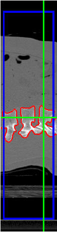 |
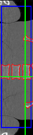
| (a) | (b) | (c) |
|
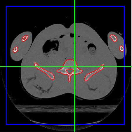
Segmentation of bones from a 512×512×125 3D CT scan of human thorax. Initial contour is in blue and final contour in red. The algorithm stopped after 392 iterations and 63 s. Segmented bones are in red, the initial surface in blue and cut planes are marked by green lines. (a) xy slice, b) xz slice c) yz slice. |
||
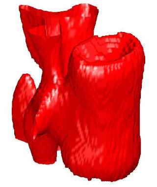 |
| 3D rendering of a segmented vertebra from a 512 × 512 × 125 CT scan. |
MRI segmentation
 |
 |
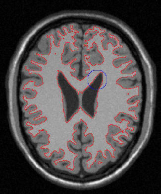
| (a) | (b) |
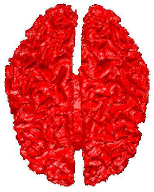
| (a) One slice of a segmented brain from a 217×217×181 3D MRI image. Initial contour is the blue circle, final contour is in red. Elapsed segmentation time was 10.52 s and 192 iterations. (b) 3D rendering of the segmented brain. | |
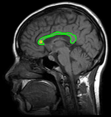 |
| Segmented corpus callosum from a 350×330 2D MRI image. The yellow circle is the initial point and the segmented corpus callosum is in green. The algorithm converged in 8 ms and 152 iterations. |
Speed comparison with traditional level sets
We have compared segmentation results and timings of the fast level set algorithm with a publicly available implementation of a standard level set algorithm in ITK library. It can be seen, that our algorithm is significantly faster then traditional level set algorithm (see the table bellow) with comparable, usually better, results.
 |
 |
| (a) | (b) |
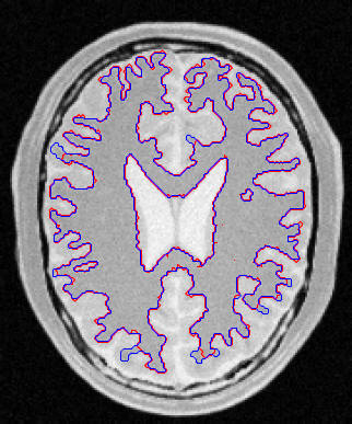
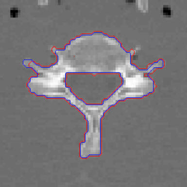
| Comparison of the fast level set method and the ITK1 threshold segmentation level set image filter results. Image (a) shows the segmentation of the gray matter from a 2D MRI image and (b) the segmentation of the cervical vertebra from a 2D CT scan. The ITK1 results are in red, the results of the fast level set method in blue. | |
|
|||||||||||||||||||||
| ITK and the fast level set timings. |
References
[1] S. Osher and J. A. Sethian, “Fronts propagation with curvature-dependent speed: algorithms based on Hamilton-Jacobi formulations,” Journal of computational physics 79, pp. 12–49, 1988.
[2] Y. Shi and W. C. Karl, “A fast level set method without solving PDEs”, IEEE International Conference on Acoustics, Speech, and Signal Processing, pp. 97-100, 2005.
[3] Jakub Krátký and Jan Kybic, „Three-dimensional segmentation of bones from CT and MRI using fast level sets“, in Proceedings of SPIE, Medical Imaging 2008: Image Processing, vol. 6914, ISBN 9780819470980, SPIE, 2008, 10 p.
[4] Jakub Krátký and Jan Kybic, „Three-dimensional segmentation of bones from CT and MRI using fast level sets“, slide presentation, April 2008.
[5] Jakub Krátký and Jan Kybic, „Introduction and application of a fast level set method for medical image segmentation“, slide presentation, June 2008.